Pseudoarthrosis or, more commonly, pseudarthrosis literally translates to “false joint.” The term Pseudarthrosis is used commonly in the situation of a fracture nonunion. For example, if the shinbone, the tibia, does not heal after a fracture, a nonunion develops. Eventually the nonunion will develop into a pseudarthrosis– related to motion between the bone ends. There is fluid and a joint like appearance to this space.
While this can happen in children, more often (but still really rare) some children are born with a pseudarthrosis, a congenital anomaly. We don’t know the reasons for these situations but these pseudoarthroses can occur in in specific locations such as the tibia. The tibia is indeed the most common location for pseudarthrosis and about 50% of the time these patients have neurofibromatosis (NF)type I. It can also occur in the clavicle although these cases are not usually related to NF- see my previous blog on this topic: http://congenitalhand.wustl.edu/2012/11/clavicle-pseudoarthrosis.html
The forearm is a common site of Pseudarthrosis in NF. This can involve the radius or, more commonly, the ulna bone. Anatomically, the forearm and the lower leg both have two bones (radius and ulna in the forearm and tibia and fibula in leg). The difficulty with a pseudoarthrosis in either location is the differential growth of the 2 bones. When one bone doesn’t grow as the other grows, deformity develops and may be accompanied by dislocation (typically of the radial head). We see similar issues in the condition of osteochondromatosis- also directly related to the differential growth. Pain can be an issue but typically is not.
 |
| Ulna pseudarthrosis. A good portion of the ulna is missing. See the forearm curve. |
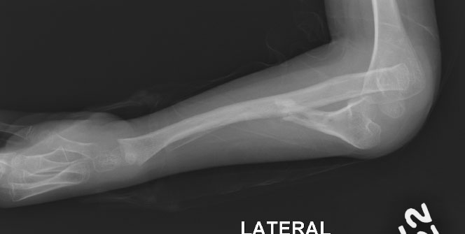 |
| Ulna pseudarthrosis from side view. |
Above is the ulna pseudoarthrosis and below is the radius pseudarthrosis.
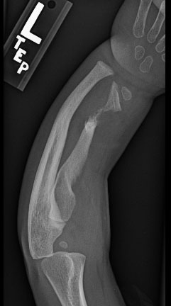 |
| Radius pseudarthrosis. Not the curvature of the forearm. The radius is very short compared to the ulna. |
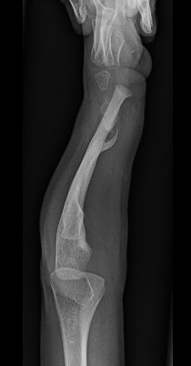 |
| Another view of radius pseudarthrosis. |
Treatment is appropriate to create a unified bone that will balance the forearm and prevent progressive deformity and angulation. There are a variety of different approaches to achieve healing and improved alignment:
1) Cleaning out the nonunion site, filling the site with bone graft (typically from hip) and often bone morphogenic protein (BMP- an off the shelf bone stimulant).
2 2) Free fibula vascularized bone transfer. In this procedure, a wider area of the Pseudarthrosissite is removed and normal bone from the fibula is substituted into the void. The beauty of this procedure is that the fibula bone is unaffected and can be brought to the forearm with a blood vessel to maximize healing. The negative of this approach is related to problem of taking this bone from the leg. While fortunately these complications are rare, they do include issues with muscle (FHL) function (affects toes), temporary sensory issues, pain, and ankle deformity.
· 3) External fixator deformity correction and bone grafting (often with BMP).