When we consider congenital abnormalities of the upper extremity, most commonly, we think of extra bones or missing digits. However, there are many other conditions which fall under this umbrella and, therefore, the care of the congenital hand surgeon. The OMT classification (Oberg, Manske, Tonkin) has recently replaced the Swanson classification as the official classification scheme for birth anomalies of the upper extremity by the IFSSH. Section III is for Dysplasias or disordered growth and includes hypertrophy, see previous post on macrodactyly and tumorous conditions. Vascular conditions including hemangiomas and malformations are thus classified.
Vascular lesions are either tumors or malformations. Hemangiomas are the most common blood vessel tumor and they are benign (i.e, not bad). Some consider these lesions a type of birthmark. Hemangiomas are one of the most common tumors of early life. These tumors are either visible at birth (congenital) or present in the first weeks or months of life (infantile). The infantile hemangiomas grow rapidly in the first 6 months to a year of life and then involute slowly (i.e., resolve). 50% have involuted by age 5, 70% by age 7 and most of the rest by age 13. There can be some hemangiomas that are visible thereafter. The congenital hemangiomas behave a bit differently as they are fully developed at birth and either disappear in the first year of life or persist.
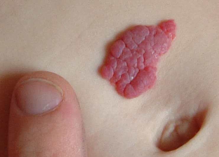
There are different types of hemangiomas including capillary, the most common in the upper extremity in my experience. These capillary hemangiomas vary in appearance but one common type is the port- wine stain. The appearance is affected by the depth of the lesion. Superficial lesions look red and slightly deeper lesions maybe bluish. Wikipedia has a good summary. Here is one capillary hemangioma from Wikipedia.
 |
| Capillary hemangioma |
Other great pictures are visible online and one good website with nice images is hemangiomaeducation
The bottom line is that most hemangiomas do not require surgical treatment as they resolve with time. However, large hemangiomas can become an appearance and social issue and family- physician discussions may be helpful.
The other type of lesion is the vascular malformations. These are more common than tumors (approximately 2:1) They are typically not visible at birth and they grow with the child to become visible or symptomatic later in life. Malformations occur when the baby is in the womb but, again, continue to grow (often very slowly) after birth and grow in proportion to the growing child. They can be venous, lymphatic, arteriovenous, capillary, etc. Venous malformations are most common.
These children may become symptomatic anytime but typically present between ages 2-5. However, I also see such children for the first time in the teenage years. Typically, neither the patient nor the family knows when the malformation appeared and it becomes progressively more bothersome as it gets larger.
Here is one example with clinical pictures of a 15 year old male with pain and an enlarging mass in the finger. It is bluish sometimes and normal colored other times. It can increase and decrease in size. This picture is, in my experience, a common situation in that the lesion does not look that “bad.” But, it is growing and can be painful. This patient and family requested surgical excision.
 |
| Venous malformation, ring finger over middle phalanx. |
Hi,
My daughter has a Hemangioma which was present at Birth March 16 measuring 2cm feeling like a fluid fild lump and now in June 16 it is measuring 5.5cm now with a blood blister starting to appear larger by the day with the fluid lump still their. She also has a larger hand (right) with bigger 3rd and 4th fingers. Is this all normal. She had an x-ray recently but still awaiting results of this. They also want to do an MRI do you think this would be a good move for a 11 week old baby girl in England?
We'd be ever so thankful for a reply if and when possible.
Our kindest regards.
Mia,
Congratulations on the birth of your daughter. Some of your daughter's symptoms are not typically seen in hemangioma. It does sound as though an MRI could provide helpful information. Good luck (I wish I could be more helpful)
Thank you ever so much. Thank you for your time in reading my comment and replying.
Hi Charles. Our four year old son developed some sort of mass in between his first and second finger on his left hand. Oxford orthopaedic Hospital think it is a haemanginoma but it seems to be growing larger and now is covering the upper part of his palm below his first finger. He doesn’t complain about any pain. He hasn’t had an mri yet only ultrasound. Should we be concerned and push for an mri? Thank you for your consideration. Kind regards. Mrs Millard.
Cotswold Mum,
Thank you for the question. An ultrasound is very helpful in making this diagnosis but an MRI is ultimately the most diagnostic test. I would certainly discuss further with your hand surgeon. I am sorry I cannot help further.