Syndactyly, simply put, means joined fingers. However, there are a number of different types including:
– cutaneous (or simple)- only a skin connection
– Complex- with a bony connection
– Complicated- with an associated syndrome
– Partial vs complete (whether part way or completely to tip of finger).
– Complex polysyndactyly. That is, extra bones and bony connections.
The partial cutanous syndactyly type is the most common followed by cutaneous complete.
The treatment philosophy for each of these syndactyly types is similar but the details and outcomes can vary. For example, we shared the results of treatment of only patient with complex syndactyly (bony connection) with this Manuscript. There has been very little shared on this topic although a variety of papers on syndactyly include a few patients that are more complex. Our goal in writing this manuscript was to focus on outcomes. And we found that these patients did not do as well as other patient types and had rotational deformities and nail abnormalities at a rate which was higher than other syndactyly patients.
We also have written about complex polysyndactyly. This type of syndactyly is even less common and we sought to provide some framework to classify these patients HERE.
I have previously written about kids with
syndactyly– those entries can be found
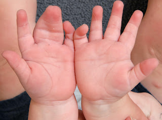
HERE.I want to briefly share images of a child with complex polysyndactyly. This child has an extra bone and joined central fingers. The thumb, index, and little finger are normal. The middle and ring finger are joined with an extra bone between.
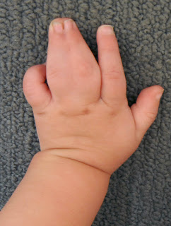 |
| Central polysyndactyly. |
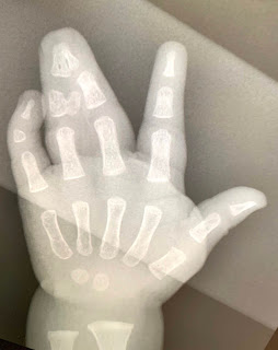 |
| Central polysyndactyly. Note the joined central bones and extra bones. |
|
 |
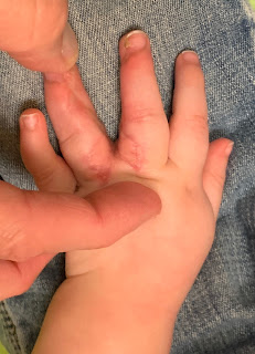
| Palm side view of central polysyndactyly. |
Syndactyly surgery aims to separate the middle and ring fingers, remove the extra bones, and reconstruct the skin to provide a long term good outcome. We hope to avoid skin creep or scarring from causing problems with the skin- specific skin incisions are chosen for this purpose. Finally, we want the appearance to be as close to ‘normal’ as possible.
In this case, we performed a syndactyly reconstruction with a skin graft substitute, a dermal substitute called hyalomatrix. This avoids the need for skin grafting. These pictures show a nice reconstruction near the hand (the commissure). The sides of the nails (the lateral nail fold) are also not quite perfect despite efforts to perfectly reconstruct that skin (we have to create that tissue). The scars are slightly prominent at 4 months after surgery but we expect that will improve with time. Finally, the hand looks great from the palm view with nice web space and alignment. The xrays also show a nice separation and bony appearance.
 |
| Hand after reconstruction of complex central polysyndactyly |
 |
| Hand after reconstruction of complex central polysyndactyly |
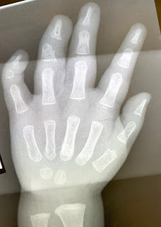 |
Xray after reconstruction of complex central polysyndactyly
|
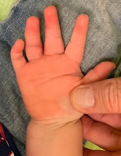 |
Hand after reconstruction of complex central polysyndactyly
|
We will continue to follow this patient until growth is done. This will help early identification of any skin tightness or creep of tissues which may occur. But, function should be excellent and the appearance should continue to get better over time.