I have posted several times on Madelungs Deformity but a recent follow- up visit with a happy patient led me to post again. One previous post was on More typical Madelungs and one on Madelungs after trauma.
Patients with an inherited Madelungs are much more common in my practice (compared to Madelungs following trauma) even though both are quite uncommon in general. It can affect one wrist or both. It can affect just the wrist or the whole forearm can be affected. Madelungs may be painful but usually the pain is mild when present. It typically affects motion of the wrist or forearm but not usually terribly. There is often visible deformity especially when looking at the wrist from the side view (see below)- it looks like the wrist “sags.” Patients typically see me as teenagers but I also see patients in their 20s and 30s- these patients are more likely to come in because of pain on the ulnar (pinky) side of the wrist.
I have am a fan of the dome osteotomy for Madelungs which was first described in Dallas. The first description Original article described the procedure and early outcome and the Second Article described longer term outcome. This paper reported very good outcomes in general but slightly less ideal outcomes in those with whole bone disease and more severe original deformity. In older patients addressing the radius bone (where the problem originates) may not be enough. In those patients, shortening the ulna bone can be an effective option as well.
Below is an 18 year old patient about 4 years after treatment for bilateral Madelungs deformity. She presented with pain, deformity, and limited range of motion. She chose to undergo surgery for her severe Madelungs. She did quite well and now has no pain, has resumed all activities, has less (but still some) deformity, and great motion. She is happy. Her x- rays are improved but still have deformity and do not look “normal.”
 |
| Madelungs deformity with classic x- ray appearance. There is a widening between the forearm bones and “v-shaped” appearance of wrist bones. |
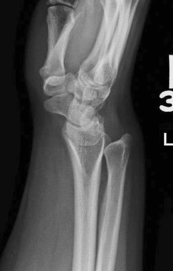 |
| Side view of Madelungs. Not the fact that the forearm bones, the radius and ulna, do not overlap. The ulna is dorsal. |
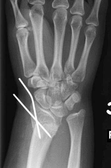 |
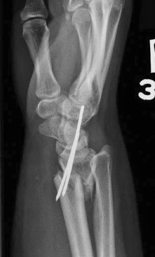
| Madelungs after osteotomy (cutting) of the radius. Metal pins in place. |
 |
| Madelungs after osteotomy, side view. |
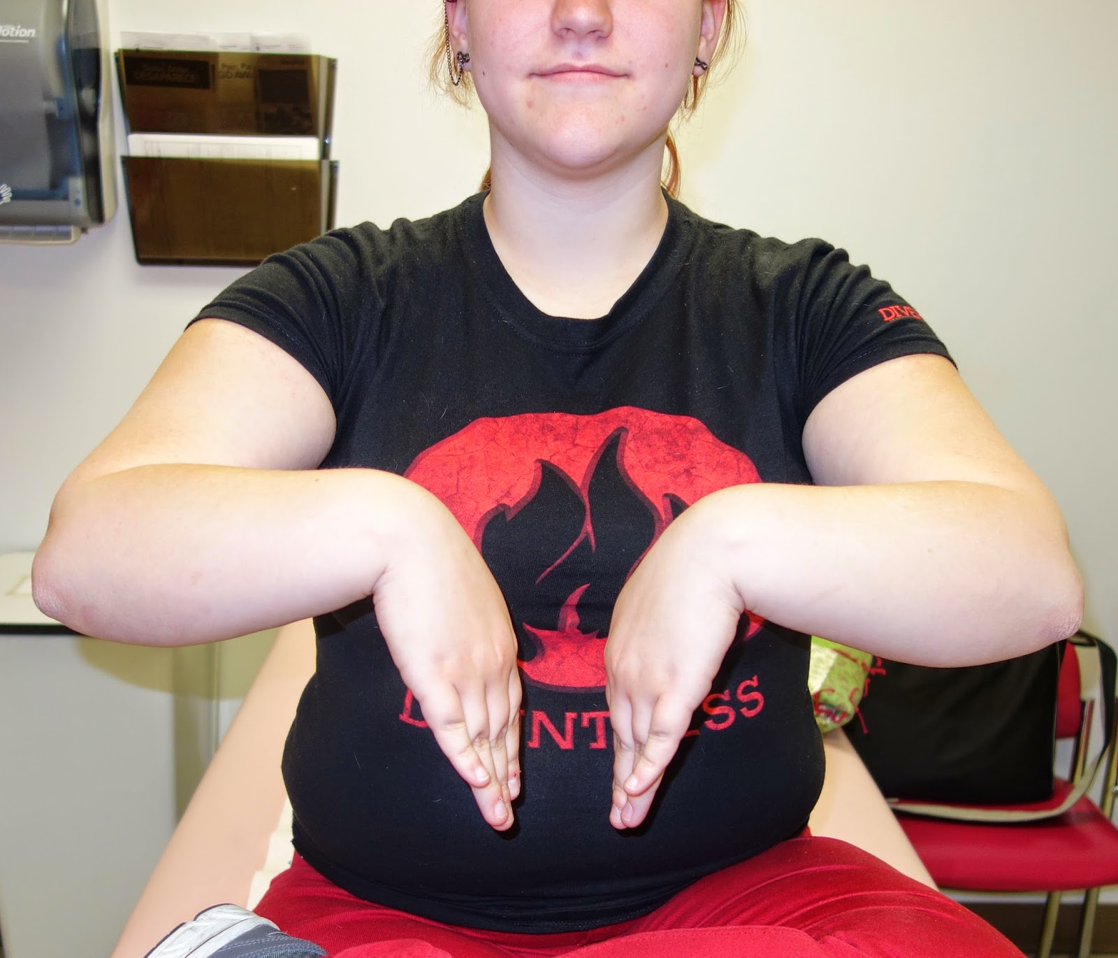
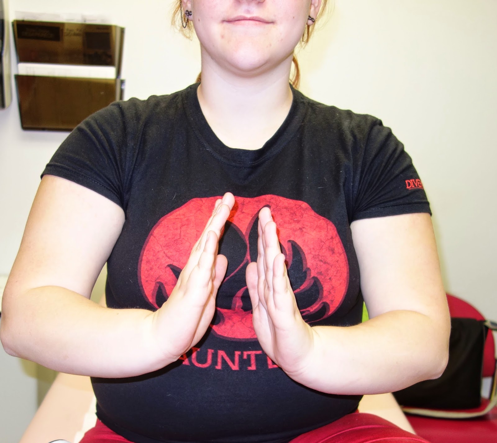
Clinical photographs demonstrating an excellent range of motion after surgery on both wrists. No pain.
 |
| Madelungs deformity after surgery. A small amount of wrist “sag” is noted. |
 |
| Madelungs final x-rays. Note appearance and shape of the bones and relationship between bones (compare to preoperative xrays above). This is not “normal” but is more anatomically correct. |
 |
| Madelungs side view with improved alignment of the bones. |