I have posted numerous times on radial longitudinal deficiency. The birth anomaly is common in my practice and I have been fortunate to meet many great kids and families with radial deficiency. A few posts can be seen HERE.
Early in my career, as influenced by my training, most patients with radial deficiency were treated with a centralization procedure. This procedure can be effective in straightening the deviated wrist but comes with a risk of the complication of shortening of an already shortened forearm (due to pressure on the growth plate of the ulna bone). Due to this concern, there is now less consensus in treatment and, at least in our practice, each child is considered individually and may be treated with a number of techniques including: observation, centralization, centralization with precentralization distraction, release with bilobed flap, and fusion. These decisions are affected by severity of the radial deficiency, the function of the fingers, the presence of a thumb, whether both arms are affected, amongst other factors.
I wanted to share somewhat early results in patient with severe radial deficiency, affected bilaterally. She had limited use of her hands due to positioning and we elected to proceed with distraction and then centralization. We used a ringed fixator (vs a uniplanar fixator) as the ringer fixator has so much more ‘power’ to correct and to do so in multiple planes. Then we centralized the wrist. The next step will be to create a thumb. Finally, we will address the other side.
 |
| Radial deficiency on the right. Note the absent radius and marked curvature of the ulna together with the deviated wrist. |
 |
| Radial deficiency on the left. Note the absent radius and marked curvature of the ulna together with the deviated wrist. |
 |
| Clinical pictures of radial deficiency, severe. Note the absent thumbs on both sides and markedly deviated wrist. |
The patient was treated with an external fixator as depicted here.
 |
| External fixator in place during distraction before centralization |
 |
| After centralization, radial deficiency wrist with temporary pin in place. Note the straight forearm/ wrist. |
 |
| After centralization, radial deficiency wrist with temporary pin in place. If you look at the middle aspect of the ulna, there is evidence of healing bone where we cut and realigned the ulna. |
|
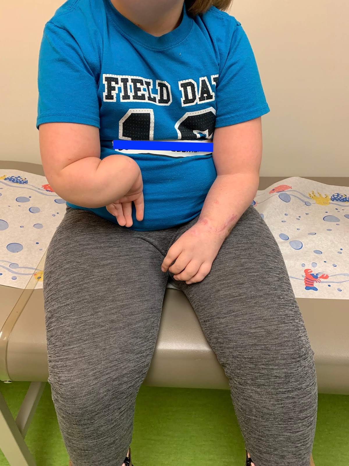
The patient is now several months after centralization. Note the difference between the two sides.
 |
| The right side with radial deficiency is untreated, the left has undergone centralization. |
 |
| The right side with radial deficiency is untreated, the left has undergone centralization. |
There is much left to offer this child and her family but we are all pleased with the initial step. Radial deficiency remains a tough challenge but there are good options to improve function and appearance.


