I have been curious about ChatGPT and so I wanted to share an encounter with artificial intelligence. Honestly, I am impressed.
The complete response:
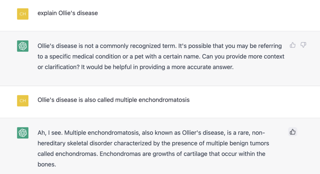
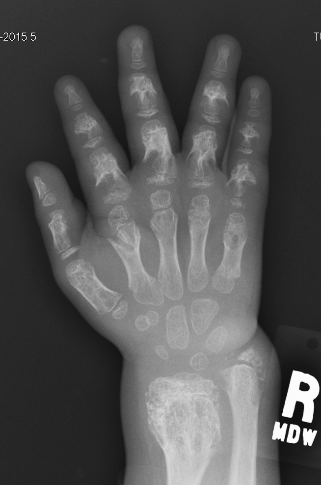
“Ah, I see. Multiple enchondromatosis, also known as Ollier’s disease, is a rare, non-hereditary skeletal disorder characterized by the presence of multiple benign tumors called enchondromas. Enchondromas are growths of cartilage that occur within the bones.
In Ollier’s disease, the enchondromas may occur in any bone, but they are most commonly found in the long bones of the arms and legs, such as the femur, tibia, humerus, and radius. The enchondromas may cause deformities or disruptions in the normal bone structure, and in severe cases, they may lead to fractures.
The exact cause of Ollier’s disease is unknown, but it is thought to be related to an abnormal growth of cells that form the cartilage in the bones. The disease usually presents itself in childhood and may progress slowly over time.
Treatment for Ollier’s disease depends on the severity of the condition and may include observation, physical therapy, and surgery. In cases where the enchondromas cause significant deformities or disrupt normal bone structure, surgical removal of the growths may be necessary to prevent fractures or other complications.”
I would add a few points to the ChatGPT response from a pediatric orthopedic viewpoint.
- Olliers is a unilateral disease with multiple enchondroma growths meaning that it affects only the right or left side of the body. Of course, this often means the upper extremity (radius and humerus) and lower extremity (femur and tibia).
2. We generally offer treatment related to bony deformity (ie, angulation) or bone shortening. These issues arise due to complete or partial growth plate arrest.
3. Treatment often includes lengthening of the bone and angular correction. There are different techniques to accomplish these goals.
Examples

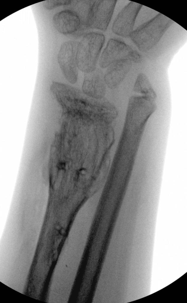
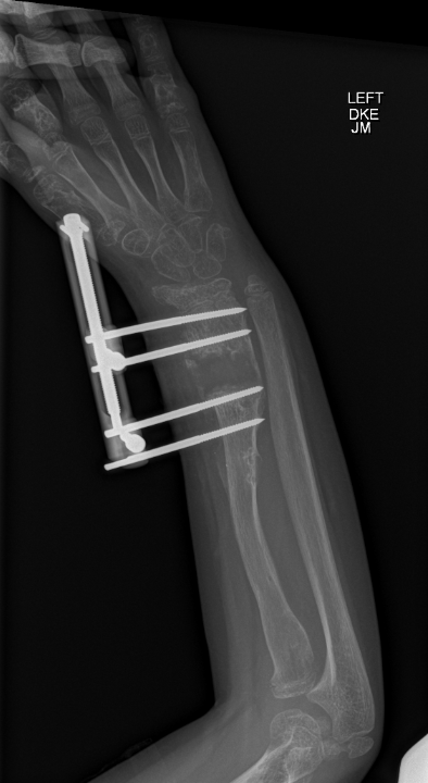
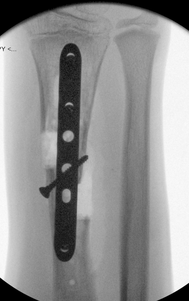
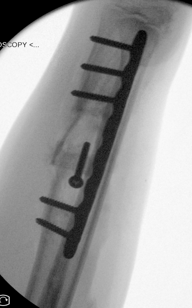
These are two techniques to lengthen the radius. The first, the step- cut, is one stage meaning that the length is gained at one surgery. The amount of lengthening is more limited and we carefully observe to avoid complications. The second technique is distraction osteogenesis. In this procedure, the first stage is cutting the bone and placing an external fixator- this can be a circular frame or a unilateral frame. We generally use a unilateral frame in these cases. 5-7 days after the surgery we start the lengthening process. This is painless and requires turning the dial 3-4 times each day to gain 0.75 to 1mm each day. Once the desired lengthening has been achieved, we leave the frame in place until the bone is solidly healed. Then, a short procedure allows removal of the fixator.
The other option, as noted above, is distraction osteogenesis. These three x-rays show the radius before the lengthening, during the lengthening, and at the time of fixator removal (ie, healed).